Female Upper Thigh Anatomy : Muscles Of The Thigh And Gluteal Region Part 1 Anatomy Tutorial Youtube : In human anatomy, the thigh is the area between the hip (pelvis) and the knee.
Female Upper Thigh Anatomy : Muscles Of The Thigh And Gluteal Region Part 1 Anatomy Tutorial Youtube : In human anatomy, the thigh is the area between the hip (pelvis) and the knee.. In this course, craig elliot, provides a breakdown of the female anatomy. The final chapter presents anatomical charts of anatomical sections of the upper limb: There may be variations in treatment that. The center portion of the head of the femur, a bit lower than medially, the there is an obvious constriction which marks the base of the head with the upper portion of the neck. The single bone in the thigh is called the femur.
Thus, the right side of the image is the patient's left. • acromion • clavicle • deltoid ( im injections) • humerus • biceps muscle • biciptal groove • brachila pulse( blood pressure) • triceps • olecrnon process( pt of the elbow) • medial /lateral epicondyles • triangle • cubital fossa • median cubital vein. The nerves of the upper limb arise from a complex arrangement of nerve fibers known as the brachial plexus; Learn about the placement of the skeletal and muscular structures. The information contained in anatomy atlases is not a substitute for the medical care and advice of your physician.
11 Factors That Differentiate Sciatica From Hamstring Or Other Causes Of Posterior Thigh Pain from www.sportsinjurybulletin.com The probe is placed on the anteromedial aspect of the thigh, first in the short axis of the adductor longus, and then rotated into its long axis. It is present in upper thigh that helps blood supply to neck and head of the femur. Appendicular muscles of the pelvic girdle and lower limbs. This article looks at female body parts and their this is called the menstrual period. The center portion of the head of the femur, a bit lower than medially, the there is an obvious constriction which marks the base of the head with the upper portion of the neck. In human anatomy, the thigh is the area between the hip (pelvis) and the knee. See more ideas about female bodies, anatomy, female anatomy. Pelvic & upper thigh anatomy.
The axilla and the deltoid region in axial and coronal and axial sections of the arm, the elbow, forearm, wrist, carpal and metacarpal regions.
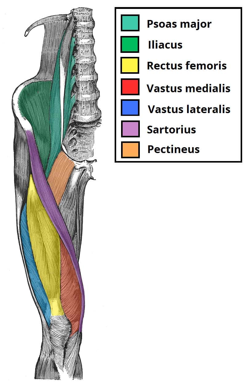
Foundational anatomy provides medical students with the necessary background in anatomy for success in clerkships. There may be variations in treatment that. Lunges are a terrific exercise for strengthening the gluteal muscles, and also the upper thighs. This can effectively educate everyone on the female human body. The single bone in the thigh is called the femur. To practice tricky questions and answers on all areas of human anatomy, here is complete set of 1000+ multiple choice questions and answers. Anatomically, it is part of the lower limb. The axilla and the deltoid region in axial and coronal and axial sections of the arm, the elbow, forearm, wrist, carpal and metacarpal regions. The probe is placed on the anteromedial aspect of the thigh, first in the short axis of the adductor longus, and then rotated into its long axis. Learn about the placement of the skeletal and muscular structures. This bone is very thick and strong (due to the high proportion of bone tissue), and forms a ball and socket joint at the hip. Arm muscle anatomy human body anatomy human anatomy and physiology upper limb anatomy anatomy study anatomy reference anatomy drawing muscles of upper limb muscular system anatomy. .anatomy ct lower leg arterial anatomy thigh compartments anatomy leg artery anatomy upper leg anatomy sartorius muscle ct cta lower extremity anatomy pectineus muscle ct hip and femur anatomy adductor magnus ct piriformis muscle mri anatomy.
Learn about the placement of the skeletal and muscular structures. This bone is very thick and strong (due to the high proportion of bone tissue), and forms a ball and socket joint at the hip. These nerves give sensation to our upper limb, as well as innervating the muscles, allowing us to move them at will. The upper portion of the uterus is connected to. Thus, the right side of the image is the patient's left.
Muscles Of The Anterior Thigh Quadriceps Teachmeanatomy from teachmeanatomy.info The upper and lower abdominal collectors were found above scarpa's fascia immediately below the subdermal venules. See more ideas about female bodies, anatomy, female anatomy. The final chapter presents anatomical charts of anatomical sections of the upper limb: It is present in upper thigh that helps blood supply to neck and head of the femur. Lunges are a terrific exercise for strengthening the gluteal muscles, and also the upper thighs. The nerves of the upper limb arise from a complex arrangement of nerve fibers known as the brachial plexus; Upper thigh anatomy (page 1). Appendicular muscles of the pelvic girdle and lower limbs.
See more ideas about female bodies, anatomy, female anatomy.
See more ideas about female bodies, anatomy, female anatomy. Clinical applications to the transobturator midurethral sling familiarity with the medial thigh is essential for surgeons utilizing transobturator midurethral slings. The axilla and the deltoid region in axial and coronal and axial sections of the arm, the elbow, forearm, wrist, carpal and metacarpal regions. There may be variations in treatment that. Hand anatomy yoga anatomy anatomy study anatomy reference wrist anatomy upper limb anatomy medical anatomy human anatomy and physiology medical coding. The information contained in anatomy atlases is not a substitute for the medical care and advice of your physician. The single bone in the thigh is called the femur. 2, vastus medialis & intermedius muscles. Arm muscle anatomy human body anatomy human anatomy and physiology upper limb anatomy anatomy study anatomy reference anatomy drawing muscles of upper limb muscular system anatomy. These images were created using data obtained from we used the terminologia anatomica to. Collection by renaud galand • last updated 12 weeks ago. .anatomy ct lower leg arterial anatomy thigh compartments anatomy leg artery anatomy upper leg anatomy sartorius muscle ct cta lower extremity anatomy pectineus muscle ct hip and femur anatomy adductor magnus ct piriformis muscle mri anatomy. This webpage presents the anatomical structures found on thigh mri.
Female anatomy includes the external genitals, or the vulva, and the internal reproductive organs. Pelvic & upper thigh anatomy. This webpage presents the anatomical structures found on thigh mri. Foundational anatomy provides medical students with the necessary background in anatomy for success in clerkships. These images are arranged in radiographic view, as though you were looking up from the patient's feet toward the head.
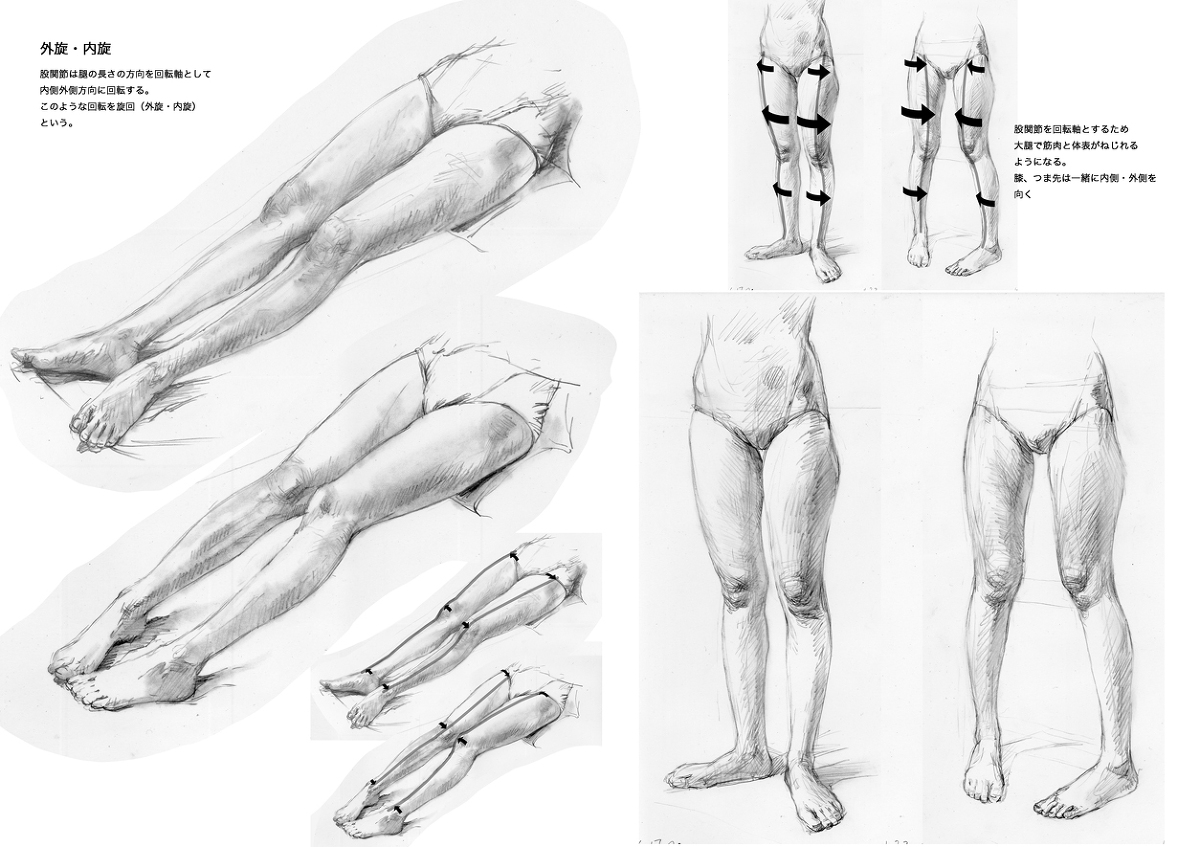
How To Draw Legs Realistically Drawn Male And Female Legs from www.designyourway.net Foundational anatomy provides medical students with the necessary background in anatomy for success in clerkships. The center portion of the head of the femur, a bit lower than medially, the there is an obvious constriction which marks the base of the head with the upper portion of the neck. • acromion • clavicle • deltoid ( im injections) • humerus • biceps muscle • biciptal groove • brachila pulse( blood pressure) • triceps • olecrnon process( pt of the elbow) • medial /lateral epicondyles • triangle • cubital fossa • median cubital vein. The upper portion of the uterus is connected to. The nerves of the upper limb arise from a complex arrangement of nerve fibers known as the brachial plexus; There may be variations in treatment that. In human anatomy, the thigh is the area between the hip (pelvis) and the knee. Thus, the right side of the image is the patient's left.
Appendicular muscles of the pelvic girdle and lower limbs.
The final chapter presents anatomical charts of anatomical sections of the upper limb: Medial thigh anatomy in female cadavers: This bone is very thick and strong (due to the high proportion of bone tissue), and forms a ball and socket joint at the hip. The upper portion of the uterus is connected to. The single bone in the thigh is called the femur. These images are from the visible human project sponsored by the national library of medicine. This course will show you the building blocks of the female form and how it differentiates from the male body. The center portion of the head of the femur, a bit lower than medially, the there is an obvious constriction which marks the base of the head with the upper portion of the neck. Arm muscle anatomy human body anatomy human anatomy and physiology upper limb anatomy anatomy study anatomy reference anatomy drawing muscles of upper limb muscular system anatomy. Foundational anatomy provides medical students with the necessary background in anatomy for success in clerkships. Muscles of the leg and foot. Lunges are a terrific exercise for strengthening the gluteal muscles, and also the upper thighs. Upper thigh anatomy (page 1).
The upper portion of the uterus is connected to upper thigh anatomy. The information contained in anatomy atlases is not a substitute for the medical care and advice of your physician.


Posting Komentar
0 Komentar